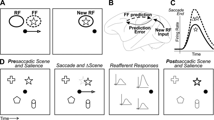
Fig. 4.
Neural-perceptual link for transsaccadic visual continuity. A: simple visual scene with 1 object, a star. Left: just before a saccade, a neuron with a receptive field (RF) as shown remaps its visual sensitivity to the future field (FF). In this example, the FF is at the star. Right: after the saccade, the presaccadic FF is replaced by the postsaccadic RF (New RF) at the same location. The response at the FF, in effect, provides a prediction of what the neuron will “see” in its New RF after the saccade. B: circuit schematic of how the FF prediction might be used. The FF prediction is sent from the FEF to extrastriate cortex (dashed arrow). It is compared with New RF input that arrives from V1 (solid arrow). Any discrepancy, or “prediction error,” is fed back to FEF and possibly used locally to modulate reafferent visual responses (dotted arrow). Modified from Crapse and Sommer (2008) with permission from Elsevier. C: the end result is that reafferent responses are tuned for transsaccadic change, as found in FEF by Crapse and Sommer (2012). The cartoon depicts reafferent responses to a star that either changed during the saccade (Δ⭐, dashed line) or stayed the same (☆, solid line). D: at the population level, the transsaccadic change-induced modulation may provide an input to saliency maps. First panel: example scene that includes the star along with other objects. For simplicity, all objects are presumed to have equal salience. Second panel: during a saccade, the star moves slightly. Third panel: after the saccade, the reafferent response across the population of neurons is enhanced for the star. Fourth panel: this increased reafferent response is read out as increased salience (or priority) of the star relative to the rest of the scene. Other influences to saliency/priority maps (e.g., attention) could be active as well but are not shown.

