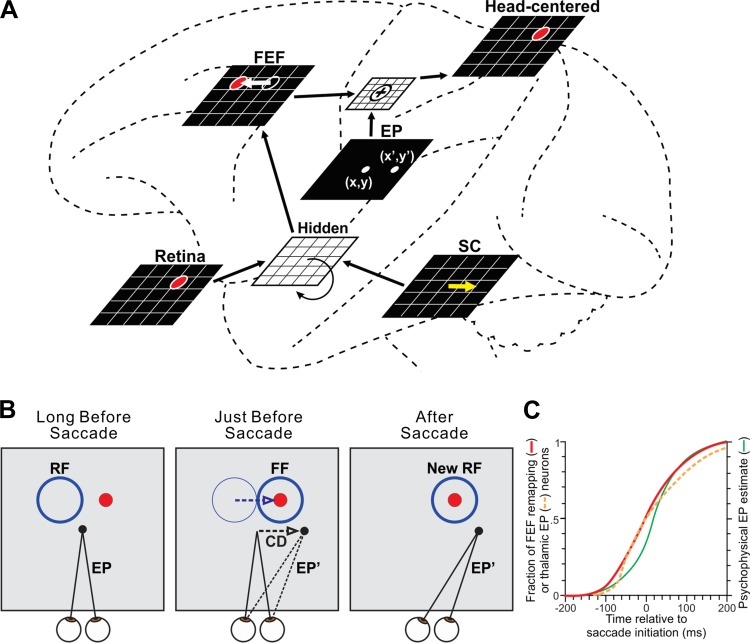
Fig. 5.
Circuit-based computational model. A: the neural network of Rao et al. (2016b), shown overlaid onto macrocircuits for remapping to illustrate concepts. Brain areas were modeled as sheets of neurons. The FEF sheet received visual and corollary discharge input mediated by a recurrent hidden layer. FEF output was combined with internal eye position (EP) signals to achieve craniotopic coordinates. The network was trained to optimize accuracy of a robot that points to a red ball (depicted as the red circle in the sheets) while making saccades. Once the model was trained, each saccade command in the SC sheet (yellow arrow) induced an equal and opposite presaccadic shift in the locus of neural activity in the FEF sheet (white arrow). B: the presaccadic shift in the FEF sheet implied a shift of sensitivity in the visual field. Consider a neuron located up and to the left in the FEF sheet (at the location of the red circle). Its classical receptive field (RF) is up and to the left. Long before the saccade, it is inactive because the red ball is not in its RF. Just before the saccade, it becomes active. It is responding to the red ball, even though the ball has not moved. The neuron's visual sensitivity has remapped to the FF. In the trained model, this remapping required corollary discharge (CD) but was time-locked to a different signal, the update in eye position from its initial location (EP) to its final location (EP′). C: evidence for synchronization of remapping and eye position updating from physiological and psychophysical studies. Shown superimposed are cumulative distributions of remapping onset times in FEF (red curve), eye position update times in thalamus (orange dashed curve), and the time course of the internal representation of eye position in humans (green curve). Based on data from Sommer and Wurtz (2006), Tanaka (2007), and Honda (1991), respectively. Modified with permission from Rao et al. (2016b). CC-BY 4.0, modified (http://journal.frontiersin.org/article/10.3389/fncom.2016.00052/full).

