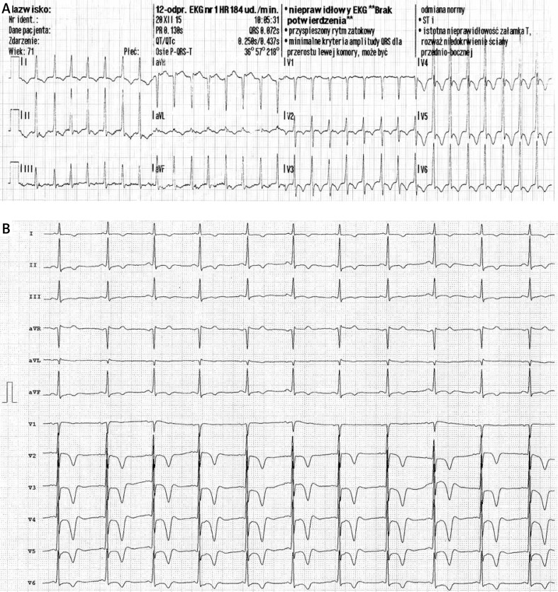
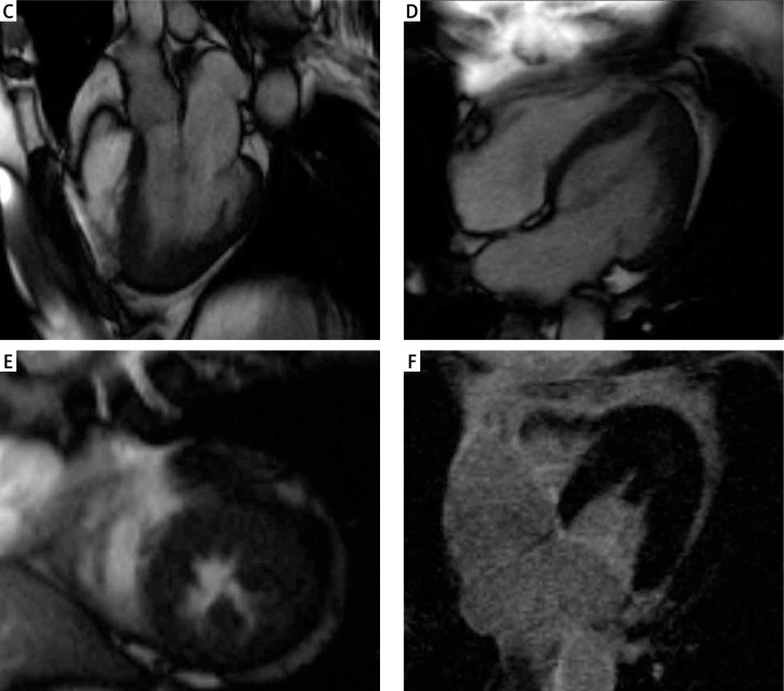
Figure 1.
A – Supraventricular tachycardia at admission with negative T waves in V2–6. B – Sinus rhythm with negative T waves in V2–6. Cine steady-state free precession cardiac magnetic resonance images in three-chamber (C) and four-chamber (D) apical long axis views as well in short axis (E) apical view. Late gadolinium enhancement imaging (F) excluded the presence of acute ischemic injury and myocardial fibrosis. Note the presence of myocardial hypertrophy in the apical segment of the lateral and anterior wall. The left ventricular ejection fraction was 83%, end-diastolic volume 79 ml, end-systolic volume 14 ml, stroke volume 65 ml, and myocardial mass 92 g


