Figure 1.
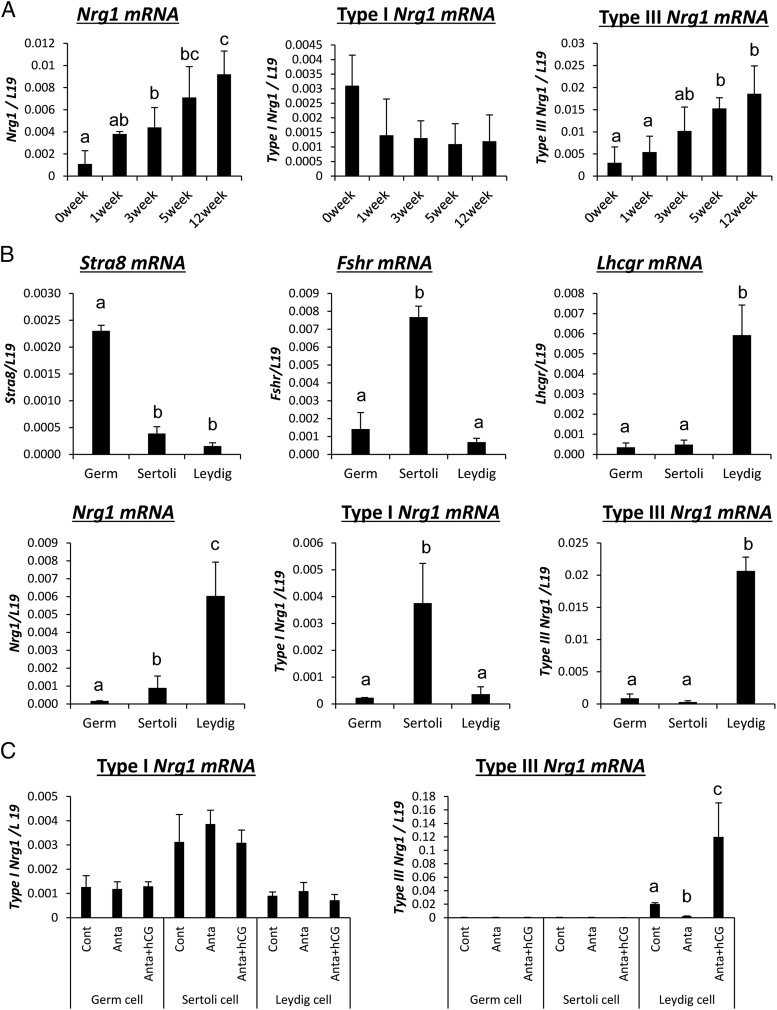
Kinetic changes of type III Nrg1 expression in LH/hCG-stimulated Leydig cells in mouse testes. A, The kinetic changes of Nrg1 (total, type I, or type III) expression in the testis collected from 0 to 12 weeks of age. Levels of mRNA were normalized to that of L19. Values are represented as the mean ± SEM of three replicates. Different superscripts denote significant differences in each cell (P < .05). B, The expression of each type of Nrg1 or specific markers of each type of testis cells in germ cells, Sertoli cells, or Leydig cells of the adult testis. Testes were collected from 12-weeks-old mice, and then percoll density gradient centrifugation was performed for the preparation of each type of cell in the testis. Levels of the gene expression were normalized to that of L19. Values are represented as the mean ± SEM of three replicates. Different superscripts denote significant differences in each cells (P < .05). C, The expression of type I Nrg1 and type III Nrg1 in each type of cell in the testis after GnRH antagonist treatment and/or hCG injection. Immature mice at 21 days of age were treated daily for 2 days with a GnRH antagonist (25 μg/d) and then received an ip injection of 10 IU hCG. Before GnRH antagonist treatment (control) and at 0 (anta) and 4 hours (hCG) after hCG, testes were collected, and then percoll density gradient centrifugation was performed for the preparation of each type of cell in the testis. The mRNA levels of type I Nrg1 and type III Nrg1 were analyzed by real-time PCR using specific primers that recognize the each specific domain. Levels of the gene expression were normalized to that of L19. Values are represented as the mean ± SEM of three replicates. Different superscripts denote significant differences in each cell (P < .05).

