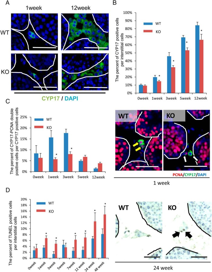
Figure 4.
The proliferation and survival of Leydig cells is suppressed in LeyNrg1KO mice. A, Immunofluorescence staining for CYP17A1, a marker of Leydig cells in testes of WT and LeyNrg1KO (KO) mice at 1 and 12 weeks of age. Scale bars correspond 100 μm. B, The percentage of CYP17A1-positive interstitial cells per total number of interstitial cells. The number of positive cells per section per testis was evaluated (n = 3 animals at each age for each genotype). Values are represented as the mean ± SEM of three sections. *, Significant differences at each age between genotypes (P < .05). C, The percentage of PCNA-CYP17A1 double-positive interstitial cells per CYP17A1-positive interstitial cells (n = 3 animals at each age for each genotype). Values are represented as the mean ± SEM of three sections. *, Significant differences at each age between genotypes (P < .05). Scale bars correspond to 5 μm. D, The percentage of TUNEL-positive interstitial cells per all of interstitial cells. The number of positive cells per section per testis was evaluated (n = 3 animals at each age for each genotype). Values are represented as the mean ± SEM of three sections. *, Significant differences at each age between genotypes (P < .05).

