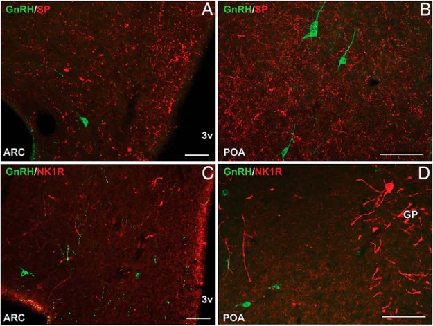
Figure 3.
Representative fluorescence photomicrographs showing lack of colocalization of GnRH (green) and SP (red) (A and B) and GnRH (green) and NK1R (red) (C and D) in the ARC (A and C) and POA (B and D). All photomicrographs are the computerized merger of 2 separate images captured sequentially with the appropriate excitation for either DyLight 488 (green) or Alexa Fluor 555 (red).Scale bar, 100 μm (A) and 50 μm (B–D). GP, globus pallidus; 3v, third ventricle.

