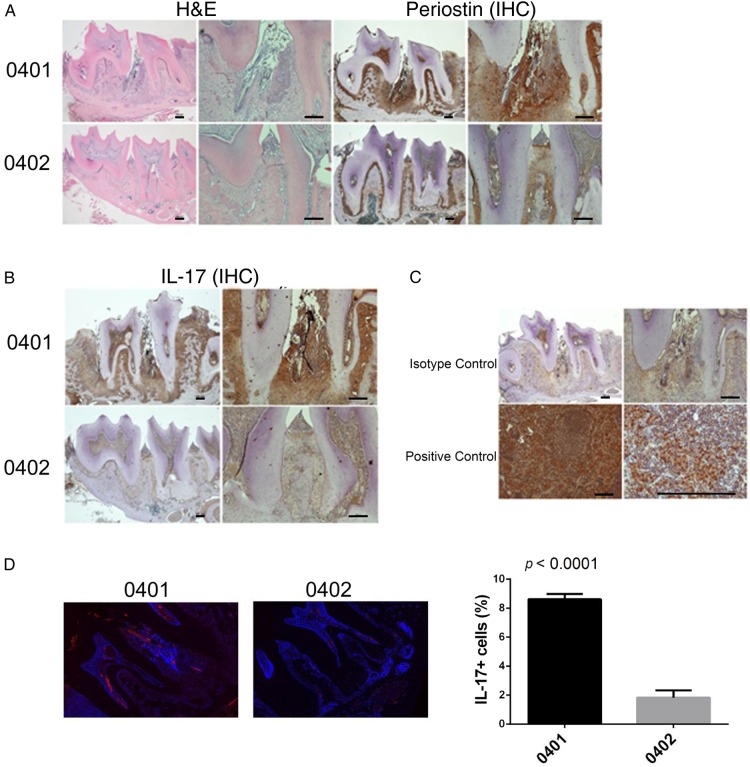
Figure 2.
Spontaneous PD in DRB1*04:01 mice. (A) Representative mandibular tissue sections stained with H&E (left) show intense inflammatory infiltrate (left). IHC staining for periostin (right) show disruption of the periodontal ligament in SE-positive, DRB1*04:01 Tg mice (top panel). SE-negative DRB1*04:02 Tg mice (bottom panel) show normal histology. (B) IHC staining for IL-17A showing markedly increased abundance of the cytokine in mandibular periodontal tissue of SE-positive, DRB1*04:01 Tg mice (top panel), compared with SE-negative, DRB1*04:02 Tg mice (bottom panel). (C) Isotype-matched antibody control shows negative IHC staining (top panel). A positive control tissue (draining lymph node—10× and 40×) shows the expected abundance of IL-17. In H&E and IHC, images of low (4×, left) and high (10×, right) magnification are shown. Horizontal bars represent 100 µm. (D) Identification of IL-17-positive cells in periodontal tissues by immunofluorescence. Red fluorescence represents IL-17; blue fluorescence (DAPI) identifies nuclei. Boxed images show higher magnification images of regions identified by white arrows. Bar graph on the right depicts mean and SEM of IL-17-positive cells in SE-positive, DRB1*04:01 Tg mice (black) and SE-negative, DRB1*04:02 Tg mice (gray), as quantified by a Biotek Cytation5 instrument. DAPI, 4', 6-diamidino-2-phenylindole; IHC, immunohistochemistry; IL, interleukin; SE, shared epitope; PD, periodontal disease.

