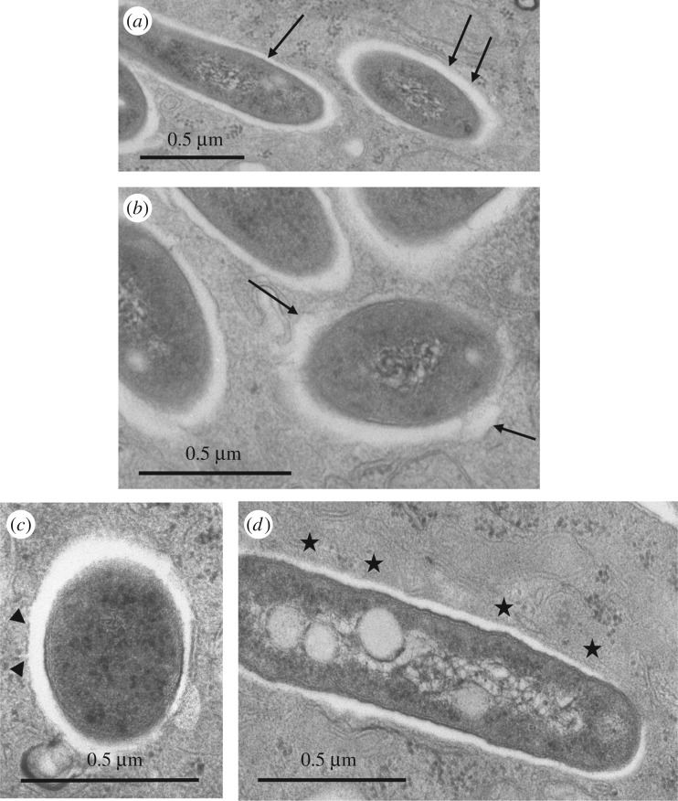
Figure 5.
Alteration of the membrane of phagosomes containing the S variant as assessed by TEM. Bone marrow-derived murine Mϕ were infected with M. abscessus S for 3 h. Phagosomes were examined for obvious signs of membrane alteration/destruction. (a) No alteration: the phagosome membrane is smooth (arrows) and closely apposed to the mycobacterial cell wall ETZ all around. (b) First sign of alteration: the phagosome membrane has become wavy and is no longer closely apposed to the bacterium all around (arrows). (c) The phagosome membrane displays breaks (arrowheads). (d) The phagosome membrane is no longer visible (stars).

