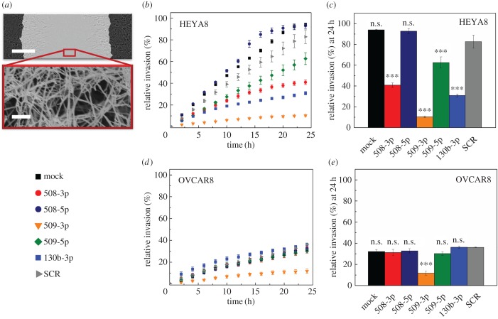
Figure 2.
Effect of miR overexpression on cell invasion through a collagen matrix. (a) Scratch wound assay where cells invade through a collagen gel. The upper panel shows a top view of the scratch; the cells on right and left sides invade into this wounded region, which is overlaid with a collagen gel. Scale bar, 200 µm. The bottom panel shows a scanning electron micrograph of the fibrous structure of a 1 mg ml−1 collagen gel. Scale bar, 1 µm. (b,d) Invasion time course for HEYA8 (N = 3) and OVCAR8 (N = 3) cells in 1 mg ml–1 collagen type I in the presence of a protease inhibitor, 50 µM GM6001. (c,e) Relative invasion of HEYA8 and OVCAR8 cells at 24 h for different miR treatments. One-way ANOVA with a Tukey post hoc test: ***p < 0.001, n.s., p > 0.05, compared to the scrambled control treatment (SCR). All error bars show the standard error of the mean.

