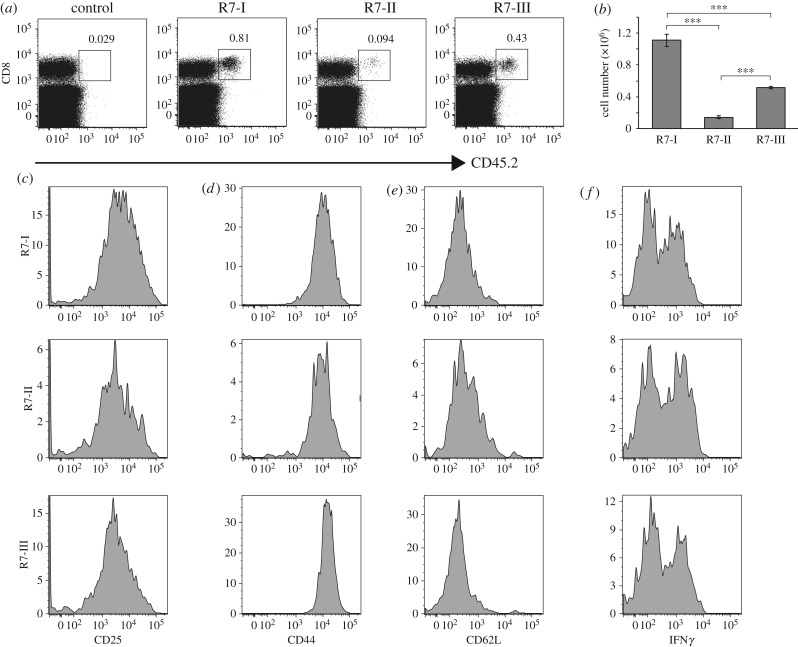
Figure 3.
Rop7-I, -II and -III CD8 T cell expansion and phenotype after Toxoplasma gondii infection. A measure of 1 × 10⁵ CD8+ tetramer+ sorted T cells from Rop7 -I, -II or -III heterozygous mice were transferred intravenously into CD45.1 congenic BALB/c mice. Twenty-four hours after T-cell transfer, mice were infected with 2 × 10⁴ Toxoplasma gondii tachyzoites. (a) Dot plots show the percentage of CD8+ CD45.2+ donor cells in the spleen at day 9 after infection. (b) Histograms show the total cell number of transferred Rop7 T cells in the spleen of recipient mice at day 9 after infection. Error bars: standard deviation (n = 3). (c–e) Histograms show CD25, CD44 and CD62 L expression on transferred Rop7 T cells in the spleen at day 9 after infection respectively. (f) Histograms show IFNγ expression by transferred Rop7 T cells from the spleen of recipient mice at day 9 after infection stimulated in vitro with Rop7 peptide. ***p < 0.001 (Student's t-test).

