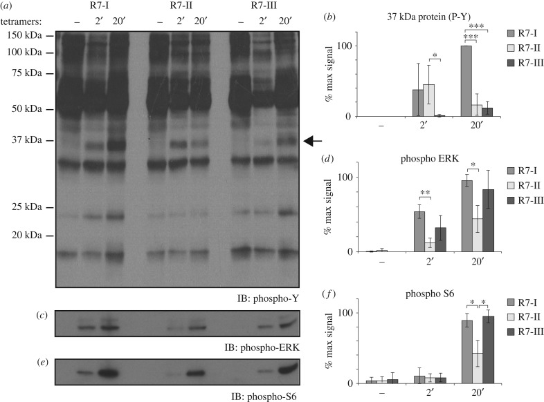
Figure 5.
TCR signalling in CD8 T cells from Rop7-I, -II and -III mice. Untouched MACS-purified CD8 T cells from pooled spleen and lymph nodes of Rop7-I, -II and -III homozygous mice were stimulated with H-2 Ld-Rop7 tetramers for 2 or 20 min. (a) Tyrosine residues, (b) ERK and (c) S6 phosphorylation was analysed in total lysates by SDS-PAGE and immunoblotting. (d–e) Quantification of band intensity for 37 kDa-size protein (arrow), phospho-ERK and phospho-S6. Average of three biological replicates. Error bars: standard deviation (n = 3). *p < 0.05, **p < 0.01, ***p < 0.001 (Student's t-test).

