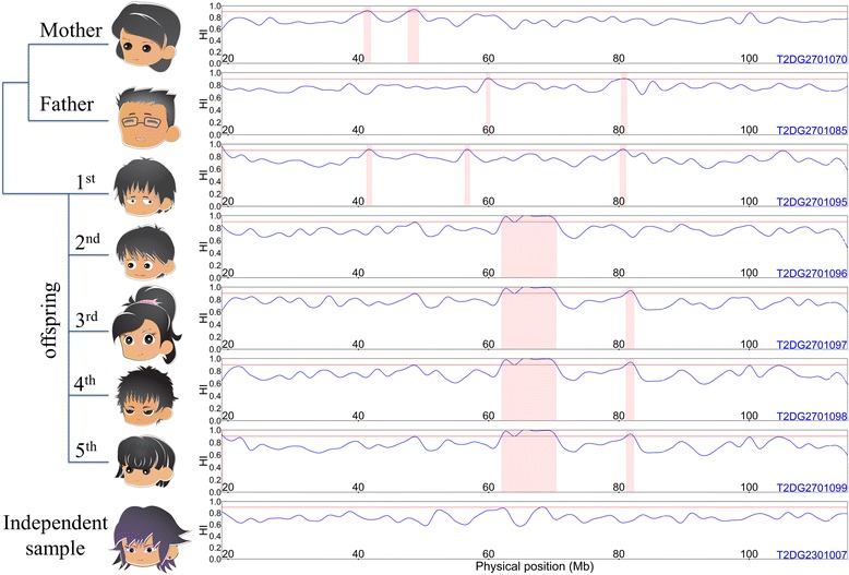
Fig. 1.

The profiling of homozygosity intensity and regions of HD on chromosome 13. The homozygosity intensity curves of seven individuals in pedigree 27 and 1 independent individual are shown. In each subfigure, the vertical axis represents homozygosity intensity (HI), which ranged from 0 to 1, and the horizontal axis represents the physical positions (Mb) of anchor SNVs of sliding windows on chromosome 13. Blue curves represent homozygosity intensity curves, and the regions shaded in pink correspond to regions of HD
