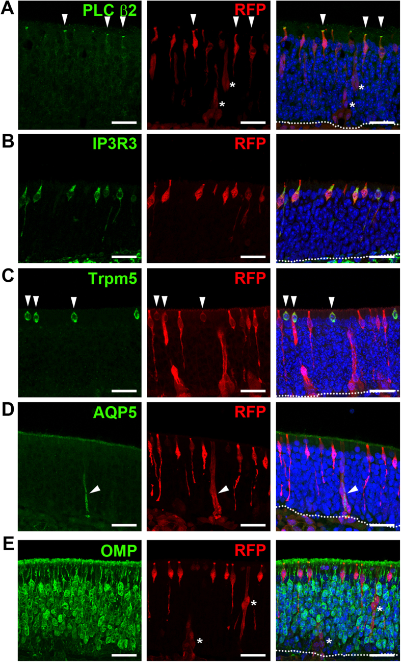
Figure 2. Ascl3-expressing cells are precursors of microvillar cells and Bowman’s glands.
Immunohistochemistry was performed on OE isolated from Ascl3EGFP-Cre/+/R26tdTomato/+ mice (2 months), using antibodies to tdTomato (RFP) and (A) PLC β2, which marks the apical microvilli of microvillar cells, (B) IP3R3, (C) Trpm5, (D) AQP5 and (E) OMP. RFP expression colocalized with microvillar cell markers: PLC β2 (arrowheads), IP3R3 and Trpm5 (arrowheads) and Bowman’s glands markers: AQP5 (arrowheads). (E) No colocalization was detected between RFP and the mature OSN marker OMP. White asterisks mark Bowman’s gland duct cells. Dotted line indicates basal lamina. Nuclei are stained by DAPI (blue). Scale bars: 25 μm.

