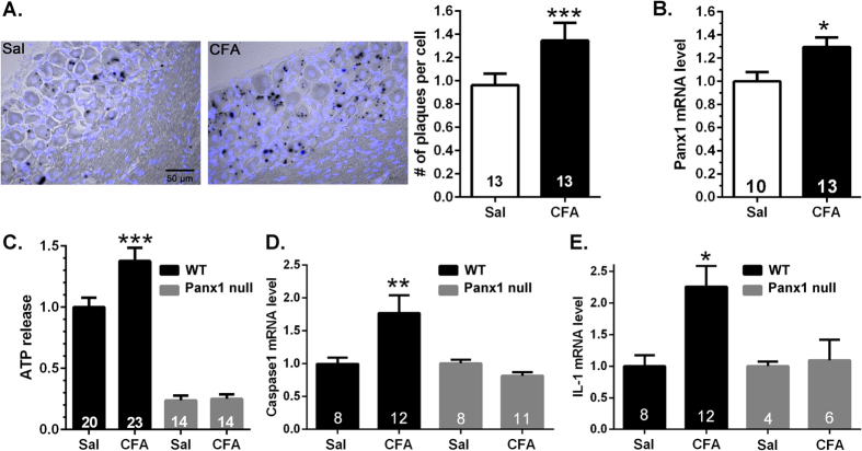
Figure 2. Increased Panx1 expression and function in trigeminal ganglia of CFA-injected mice.
Histograms showing the mean ± SE values of the (A) number of LacZ plaques per cell (satellite glia and sensory neurons) and of (B) Panx1 mRNA levels in the trigeminal ganglia of mice 7 days after submandibular injection of CFA relative to those obtained from saline (Sal) injected ones. Numbers of mice are indicated in bars. *P < 0.05, **P < 0.01, unpaired t-test. (C–E) ATP release and caspase-1 and IL-1β mRNA levels are elevated in trigeminal ganglia following one week CFA injection and these changes are prevented in mice lacking Panx1. *P < 0.05, **P < 0.01, ***P < 0.001, one way ANOVA followed by Tukeys’ multiple comparison test. Numbers of mice are indicated in bars.

