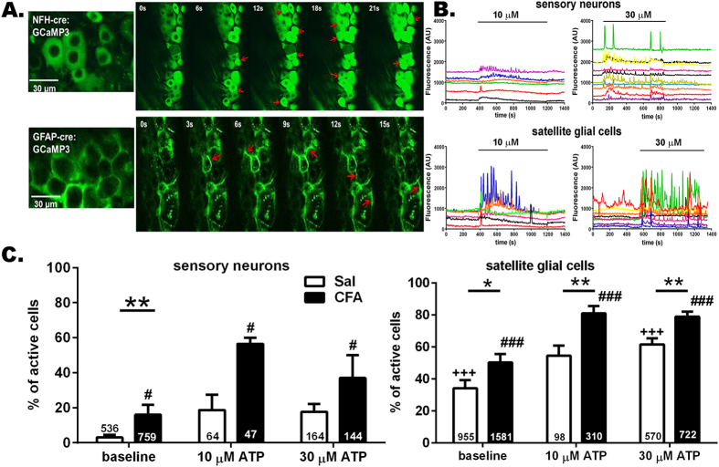
Figure 3. ATP enhances hyperactivity of trigeminal ganglion neurons and satellite glia of mice with orofacial pain.
(A) Confocal images showing GCaMP3 fluorescence changes in intact trigeminal ganglia reporting Ca2+ dynamics in neurons with NFH-Cre:GCaMP3 expression and satellite glial cells with GFAP-Cre:GCaMP3 expression. Red arrows indicate representative neurons and satellite glial cells showing Ca2+ elevations during this short (21 and 15 sec in upper and lower panels) recording epochs. (B) ATP stimulation of trigeminal ganglia increased intracellular Ca2+ concentration in sensory neurons and satellite glial cells. Representative traces of Ca2+ transients recorded from neurons and satellite glial cells to increasing concentrations of ATP (10 and 30 μM). (C) The proportion of active neurons and satellite glial cells at baseline is significantly higher in trigeminal ganglia from CFA-injected than in saline-injected mice; application of ATP increased the proportion of active neurons and satellite glial cells in trigeminal ganglia derived from one week CFA-treated mice (baseline vs ATP). *P < 0.05, **P < 0.01, one way ANOVA (saline vs CFA). +++P < 0.005, #P < 0.05 and ###P < 0.001, one way ANOVA followed by Sidak’s multiple comparison test for ATP doses in saline and CFA groups, respectively. The total number of cells analyzed derived from 4-15 mice are indicated in the bars.

