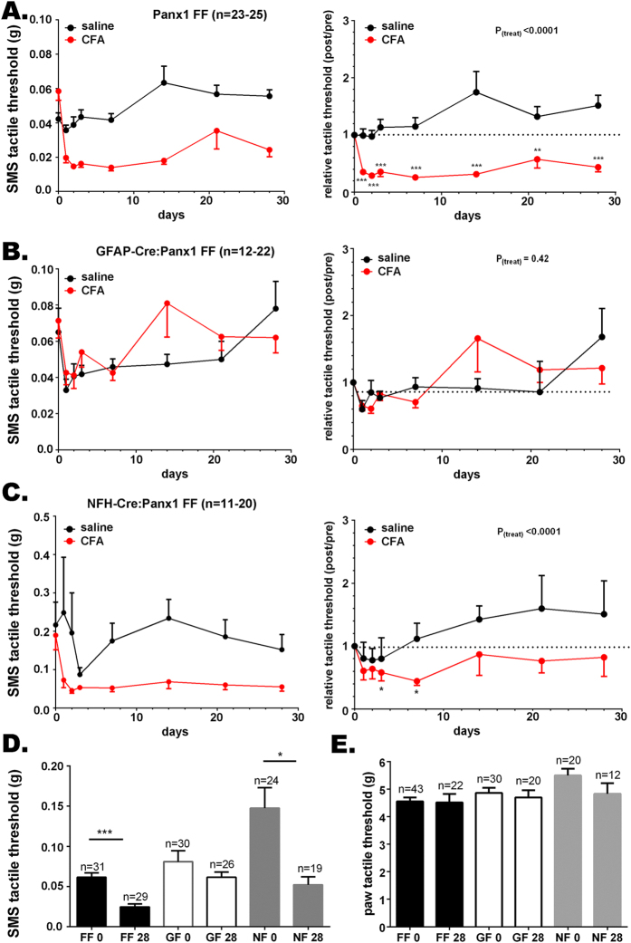
Figure 4. Panx1 deletion in GFAP-positive glia and neurons differentially affects tactile hypersensitivity.
Time course of changes of submandibular skin (SMS) tactile threshold of (A left) Panx1fl/fl, (B left) GFAP-Cre:Panx1fl/fl, and (C left) NFH-Cre:Panx1fl/fl following CFA injection into the submandibular skin; (A–C right) are the normalized values (post/pre-CFA injection) of SMS tactile threshold shown in part A. Histograms of the means ± SE values of SMS (D) and hind paw (E) tactile threshold obtained before (0 days) and at 28 days post-CFA injection into the submandibular skin. Parts A–C: *P < 0.05, **P < 0.01, ***P < 0.001 from one-way ANOVA with repeated measures. Parts D-E: *P < 0.05, ***P < 0.001, One-way ANOVA followed by Tukeys’ multiple comparison tests. Number of mice indicated. FF: Panx1fl/fl, GF: GFAP-Cre:Panx1fl/fl; NF: NFH-Cre:Panx1fl/fl; 0 and 28 indexes: before and 28 days after CFA injection, respectively.

