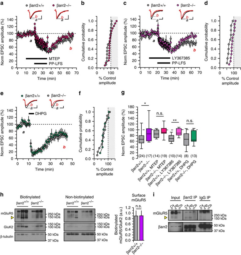
Figure 6. mGluR5 is necessary for LTD induced by synaptic stimulation.
(a,b) Inhibition of mGluR5 by MTEP (1 μM) attenuates prolonged Sch-CA1 synaptic depression in βarr2+/+ and βarr2−/− mice. A time course plot shows depression of EPSC amplitudes during PP-LFS in the presence of MTEP, but normalized amplitudes in both βarr2+/+ and βarr2−/− animals recover by 30 min after PP-LFS (P=0.27, n=14 cells from seven βarr2+/+ mice, n=19 cells from 12 βarr2−/− mice). Every third event was plotted during basal stimulation, and every ninth event was plotted during PP-LFS. A cumulative histogram further summarizes post-train data. (c,d) Using LY367385 (50 μM), an mGluR1 antagonist, PP-LFS LTD is observed in βarr2+/+, but not βarr2−/− mice. A time course plot illustrates depression of EPSCs in βarr2+/+ following PP-LFS in the presence of LY367385 and recovery to baseline levels in βarr2−/− animals (P=0.003, n=10 cells from six βarr2+/+ mice, n=14 cells from eight βarr2−/− mice); data is summarized in a cumulative histogram. (e,f) DHPG induces LTD in βarr2+/+ and βarr2−/− mice. βarr2+/+, and βarr2−/− recordings have similar time courses for depression and post-train amplitude distributions (P=0.86, n=13 cells from six wild-type mice, n=8 cells from four knockout mice). (g) A box plot shows recovery of all βarr2−/− conditions from PP-LFS LTD but not DHPG LTD; PP-LFS LTD is abrogated in βarr2+/+ recordings only by MTEP. (h) Monomeric mGluR5 (yellow arrow) and GluK2 immunoreactivity was detected in both the βarr2+/+ and βarr2−/− surface biotinylated fractions by western blotting, and less so in non-biotinylated fractions (n=3 pairs of βarr2+/+ and βarr2−/− mice). β-tubulin immunoreactivity indicated that intracellular proteins were enriched in the non-biotinylated fraction. Quantitated monomeric mGluR5 optical density normalized against the GluK2 signal is provided in a bar graph illustrating the mean and s.e.m. (i) mGluR5 co-immunoprecipitates with β-arrestin2 in the cortex and hippocampus of wild-type mouse brains (n=5 blots). Pull-down by an IgG isotype antibody yields little mGlu5 receptor immunoreactivity.β-arrestin2 is strongly detected in the immunoprecipitation and input conditions. Representative traces calibration: x axes, 10 ms; y axes, 100 pA. Groups were compared by Mann–Whitney tests. Asterisks denote significance (*P<0.05, **P<0.01); n.s., non-significant.

