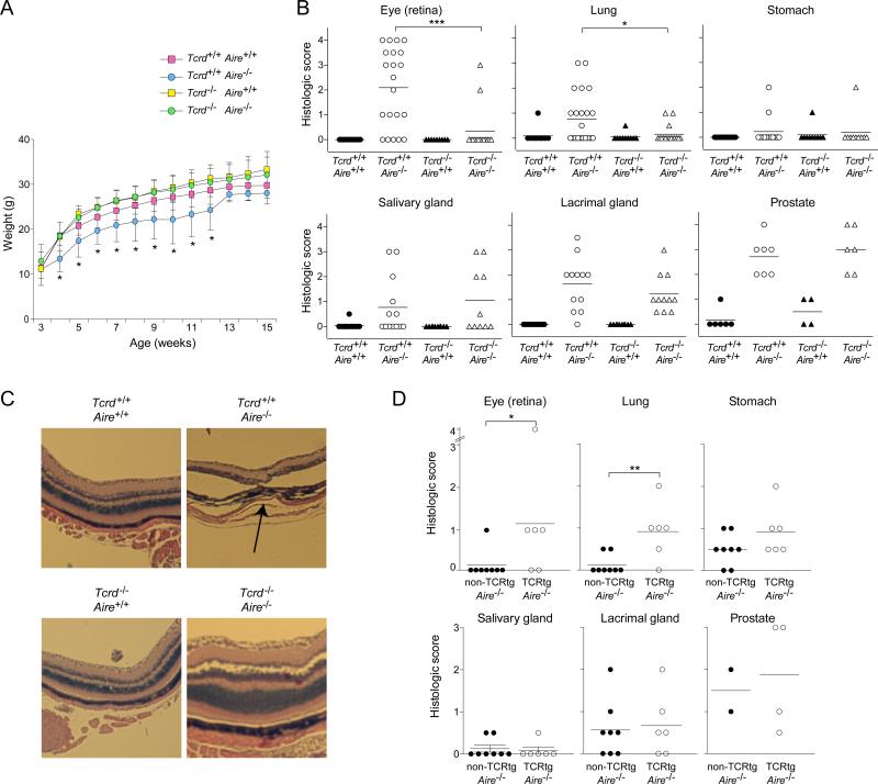
Fig 5. γδT cells played a role in the autoimmune disease characteristic of Aire-deficient mice.
Littermates carrying a homozygous null mutation of Aire, Tcrd, both or neither on the B6 genetic background were followed until 15 weeks of age, when the indicated organs were taken for histology (n=10 per group). A. Weight curves. B. Histologic scores, employing the scoring system described in Experimental Procedures. Statistics as per Fig. 1. C. Eye histology. Hematoxylin and eosin staining of retinal tissue. Arrow indicates the disrupted retinal layer. D. As per panel B, except mice harboring a homozygous null-mutation of Aire that either did or did not carry a Vγ6+Vδ1+ TCR transgene were followed until 12 weeks of age (n=2-4 for prostate, 6-8 for all other tissues). Statistics as per Fig. 1. (See also Fig. S2)

