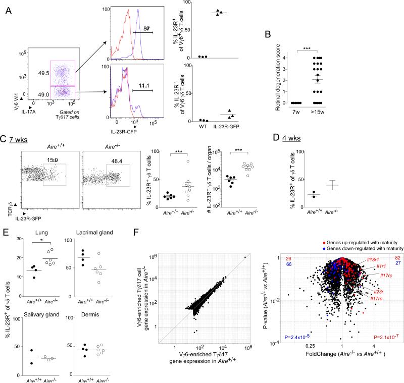
Fig. 6. Tγδ17 cells accumulated in the retina prior to autoimmune attack in Aire-deficient mice.
A. IL-23 reporter validation. Flow cytometric analysis of thymic Tγδ17 cells from day-of-birth IL-23R-GFP mice. Left and center panels, typical cytofluorometric plots; right panel, summary data (n=3). B. Histologic scores, as per Experimental Procedures, of eye tissue from Aire−/− mice taken at 7 or 15 weeks of age (n=6-20). C. Cytoflurometric comparison of GFP+ Tγδ17 cells from uveoretinal tissue of 7-week-old Aire+/+ and Aire−/− mice carrying the IL-23R-GFP reporter. Left, typical profiles; right, summary data on fractional representation and number (n=6-8). D. Summary data for 4-week-old mice (n=2-3). E. As per panel C, except cells were isolated from lung, lacrimal gland, or dermis (n=2-3 for salivary gland, 4-6 for all other tissues). F. RNA-seq analysis of gene expression in Vγ1,2,4,5− CD27− (i.e. IL-17+ Vγ6+-enriched) γδ T cells isolated from the lungs of 10-week-old Aire+/+ or Aire−/− mice (n=2). Red- or blue-highlighted genes represent loci two-fold up- or down-regulated during Vγ6+ cell thymic maturation (Narayan et al., 2012), respectively. p-values reflect the significance of signature enrichment as calculated by the χ2 -test. Other statistics as per Fig. 1. (See also Fig. S3). Dot plot scales for this figure are all the same.

