
Figure 1.
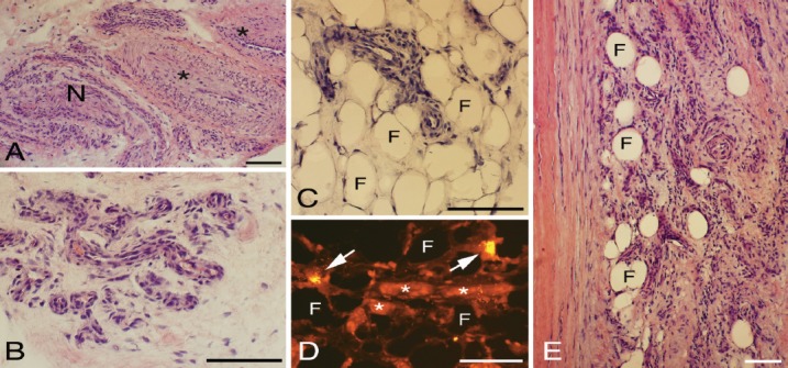
Peritendinous tissue located close to the plantaris tendon and where this tendon is facing the Achilles tendon. Staining for H&E in (A-C and E). Staining with antibody against mast cell tryptase in (D). There is a presence of large arterioles (asterisks, A) and numerous small vessels (B,E). The small vessels are partially embedded within the here occurring fat tissue (asterisks, C-E). Also in (D), there is a presence of fine vessels. Tendon tissue proper to the left in (E). Arrows in (D) point at mast cells. F= fat tissue (C-E); N= nerve fascicle (A). Bars=100 μm.