
Figure 2.
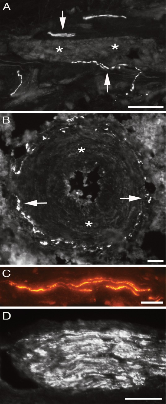
Peritendinous connective tissue in between Achilles and plantaris tendons showing PGP9.5 positive immunoreactions (arrows) in close vicinity of a longitudinally/obliqually cut blood vessel (A), in the media-adventitia junction of the wall of a large arteriole (B), in the form of fine isolated nerve fibres (C) and in a nerve fascicle (D). Asterisks in the media of the blood vessel walls. Bars=40 μm.