
Figure 6.
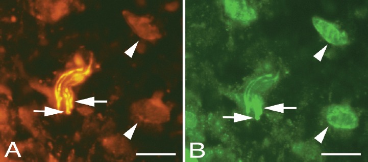
Peritendinous connective tissue in between Achilles and plantaris tendons double stained for PGP9.5 (yellow, A) and NMDAR1 (green/white, B). Overlapping positive stainings are seen in some nerve fibres (arrows). Cells in the peritendinous tissue (arrowheads) are seen to exhibit NMDAR1 immunoreaction, but only unspecific staining in the PGP9.5 stained section. Bars=20 μm.