
Figure 7.
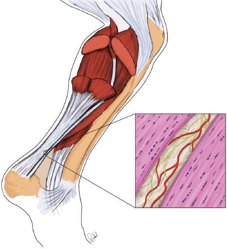
Schematic drawing showing the highly innervated and vascularised peritendinous connective tissue in between the plantaris and Achilles tendons, both showing tendinosis features such as tenocytes frequently being lined up in rows. Red marks vessels, yellow innervation.