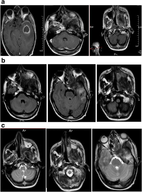
Fig. 1.

Magnetic resonance imaging studies show a tumor process of the skull base involving the sphenoid bone with its two left wings, the squamous part of the left temporal bone, associated with intracranial expansion, as well as a second, isolated mass in the temporal lobe. a T1-weighted, gadolinium-enhanced axial magnetic resonance imaging scans. b T1-weighted, axial magnetic resonance imaging scans. c T2-weighted axial magnetic resonance imaging scans
