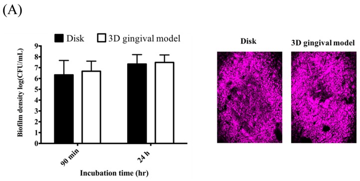
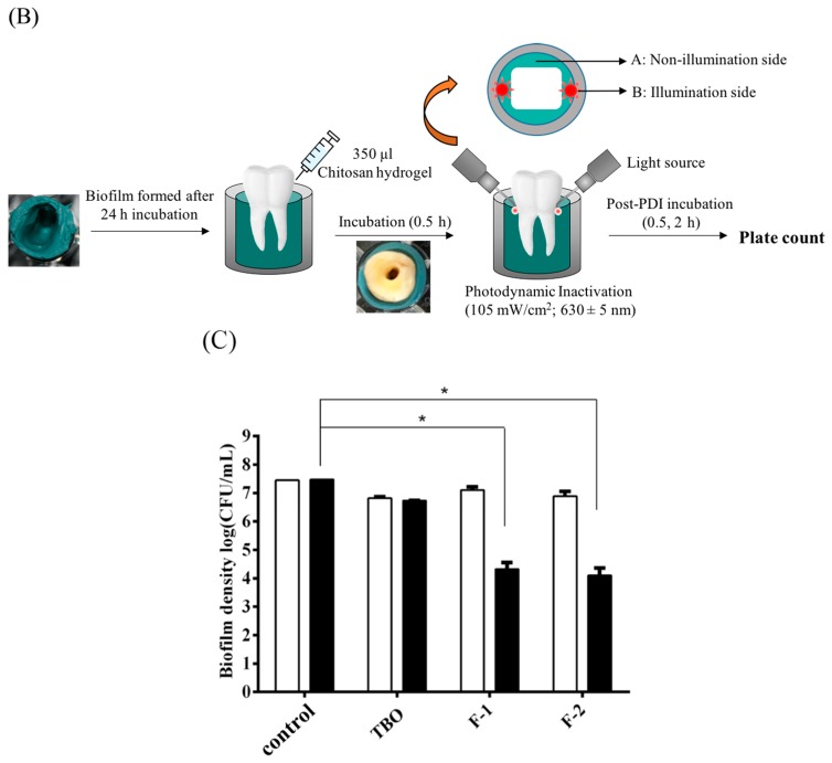
Figure 1.
(A) Comparison of the biofilm density (left panel) and extracellular polymeric substance (right panel) of S. aureus biofilms cultured in the disk model and 3D gingival model, respectively; (B) Schematic illustration of the procedures of chitosan hydrogel mediated photodynamic inactivation (PDI) in 3D gingival model; (C) Cell survival fraction of S. aureus biofilm in 3D gingival model treated will chitosan hydrogel-mediated PDI. Biofilm cells were incubated with 20 μM TBO or chitosan hydrogels containing 20 μM toluidine blue O (TBO) for 0.5 h, followed by light exposure on two sides of the 3D gingival model (irradiation dose: 5.4 J·cm−2 for each side; total light dose: 10.8 J·cm−2). After PDI, S. aureus biofilm was maintained in contact with the chitosan hydrogels with an additional 0.5 h (☐) and 2 h (■) incubation prior to plate count. Each value is the mean of three independent experiments ± standard deviation. * p < 0.05.


