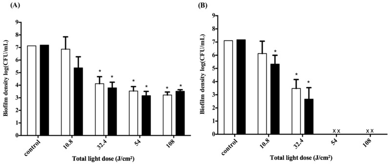
Figure 3.
Cell survival fraction of S. aureus biofilm in 3D gingival model after incubation with F-2 for 0.5 h, and subjected to various doses of light illumination from two sides. After PDI, S. aureus biofilm was maintained in contact with F-2 for 0.5 h (☐) and 2 h (■) prior to plate count. (A) Non-illumination side; (B) illumination side. The locations of (A) and (B) are shown in Figure 1B. Each value is the mean of three independent experiments ± standard deviation. * p < 0.05.

