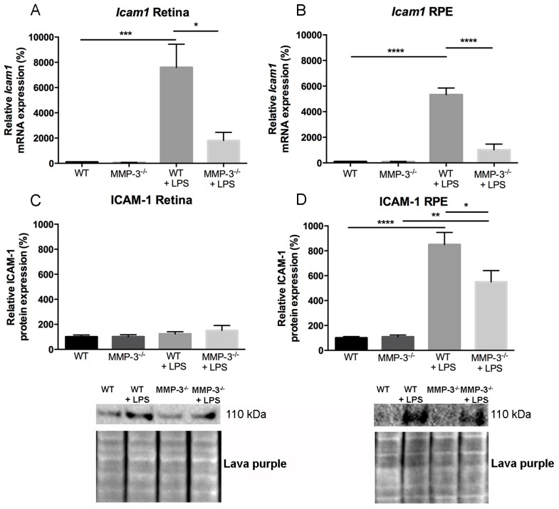
Figure 6.
The effect of MMP-3 deficiency on ICAM-1 mRNA and protein levels in the retina and RPE during EIU. (A,B) Real time-PCR data revealed significantly elevated levels of Icam1 at 16 h after EIU induction in the posterior eye segment of WT eyes. This augmentation was significantly reduced in retinal and RPE samples of MMP-3−/− mice as compared to WT animals. Data were normalized against the reference genes and were expressed as a percentage relative to 0 hpi (n ≥ 3); (C) The protein level of ICAM-1 in the retina only showed a slight increase after EIU, and no clear difference between WT and MMP-3−/− mice after LPS; (D) However, ICAM-1 in the RPE was significantly increased at 16 hpi in both genotypes. Interestingly, MMP-3 deficiency reduced ICAM-1 levels after LPS. Lava purple staining served as a loading control. Western blot data were normalized against Lava purple total protein stain and expressed as a percentage relative to 0 hpi. All data are shown as mean ± SEM, n ≥ 5, * p ≤ 0.05, ** p ≤ 0.01, *** p ≤ 0.001, **** p ≤ 0.0001. Data with no indicated statistical analysis point out no significant results.

