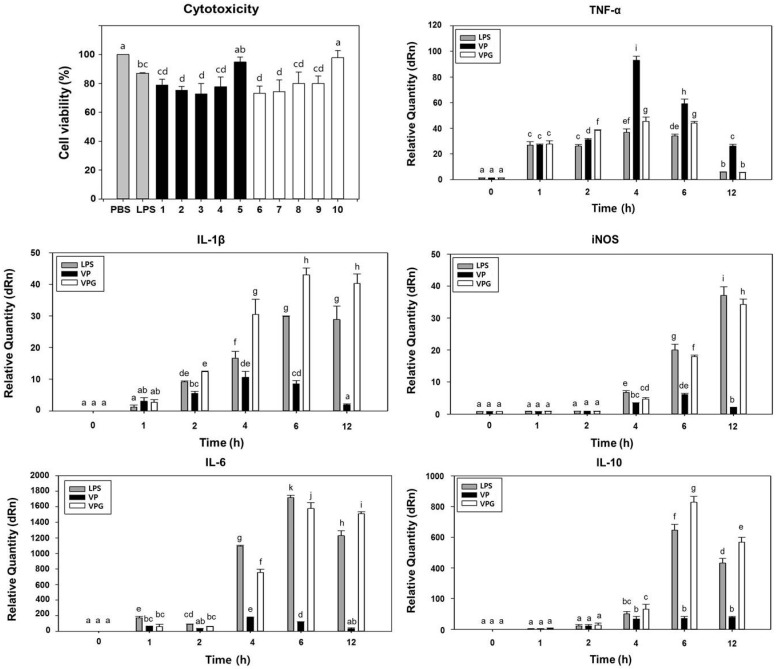
Figure 3.
VPGs-exposed murine macrophage (RAW 264.7) shows cell viability and stimulates both pro- and anti-inflammatory cytokine production. To determine cytotoxicity, macrophages were exposed to PBS buffer, LPS from Escherichia coli, V. parahaemolyticus wild-type cells (bars 1–5), and VPGs treated with NaOH for 60 min (bars 6–10), respectively. At 24 h post-exposure, macrophages were collected for analysis of cell viability using Cell counting Kit-8. Bars represent exposure doses of 2.5 × 10⁶ (1 and 6), 1.7 × 10⁶ (2 and 7), 1.3 × 10⁶ (3 and 8), 1.0 × 10⁶ (4 and 9), and 0.5 × 10⁶ (5 and 10) CFU/mL, respectively. Absorbance was measured at 450 nm and all experiments were performed in triplicate. Cytotoxic activity is expressed as the percentage of cell viability by the formula described in Materials and Methods. At 4 h post-exposure with LPS from Escherichia coli, V. parahaemolyticus wild-type cells (VP), and NaOH-induced VPGs for 60 min, respectively, macrophages were collected for analysis of gene expression for cytokines TNF-α; IL-1β; iNOS; IL-6; and IL-10 using RT-qPCR. Data are representative of triplicate experiments with each sample run in triplicate. All data are expressed as the mean ± the standard error of the mean. Mean separation by Duncan’s multiple range test at p < 0.05. The same letter above bars represented no significant difference between treatments.

