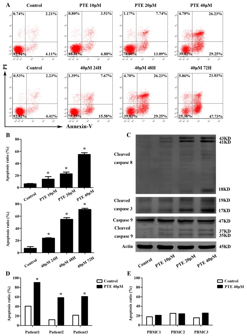
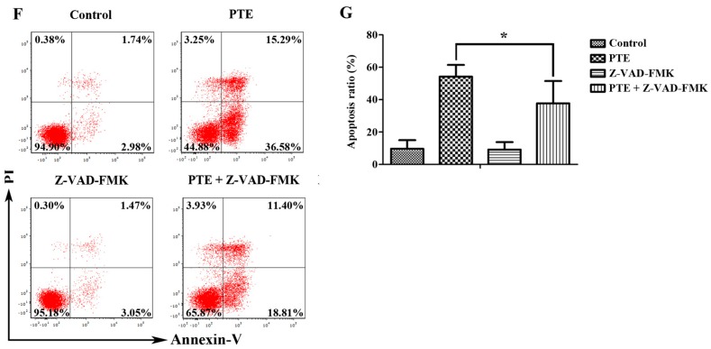
Figure 2.
PTE enhances the apoptosis of H929 cells, primary CD138+ MM cells and caspase activation. (A) H929 cells were treated with PTE (10, 20, and 40 μM) for 48 h or treated with 40 μM for 24, 48 and 72 h, stained with AnnexinV- fluorescein isothiocyanate (FITC) / propidium iodide (PI) and analyzed by flow cytometry; (B) the percentage of FITC positive cells treated with 0, 10, 20, and 40 μM of PTE. Data is presented as mean ± SD (n = 3, * p < 0.05); (C) the protein levels of cleaved caspase-3, cleaved capase-8 and caspase-9 were determined by Western blot; (D,E) primary CD138+ MM cells and peripheral blood mononuclear cells (PBMCs) were treated with 40 μM of PTE for 48 h stained with AnnexinV-FITC/PI and analyzed by flow cytometry; (F) H929 cells were pre-incubated with or without Z-VAD-FMK (50 μM) for 3 h and then treated with PTE (40 μM) for 48 h, stained with AnnexinV-FITC/PI and analyzed by flow cytometry; and (G) the percentage of FITC positive cells treated with 40 μM of PTE that pre-incubated with or without Z-VAD-FMK. Data is presented as mean ± SD (n = 3, * p < 0.05).


