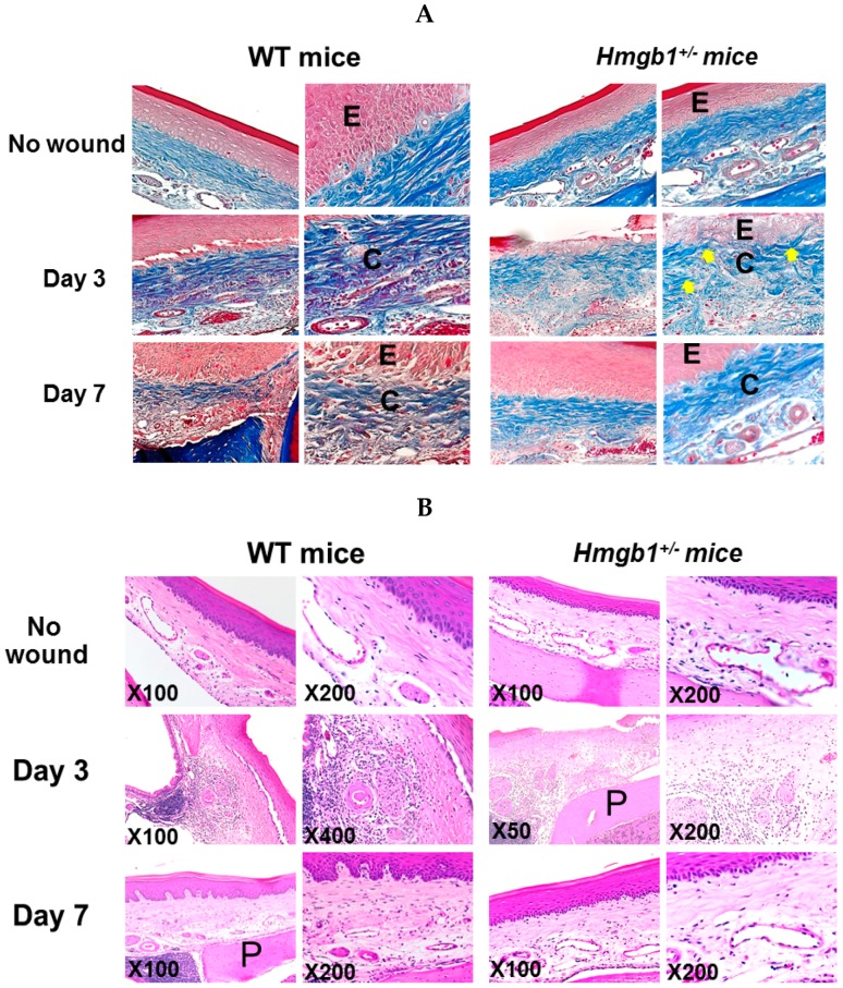
Figure 3.
Reduction of collagen fibers and delayed re-epithelialization in Hmgb1+/− wound on Day 3 after wound surgery. (A) Sections were stained with Mallory’s azan. Representative palatal wound sections show the collagen bundles of WT and Hmgb1+/− mice. Note that blue indicates collagen bundle stained by Mallory’s azan stain. Arrows indicate weave-like pattern of collagen bundles in Hmgb1+/− mice. Original magnifications: left panels 200×, right panels 400×; (B) Sections were stained with hematoxylin and eosin. E, epithelium; C, collagen bundle; P, palatal bone. n = 5 wounds for each time point and genotype.

