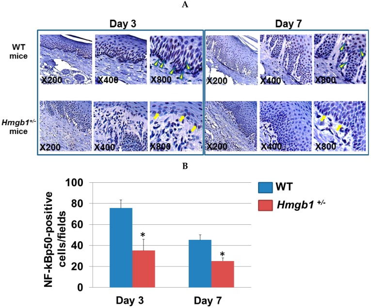
Figure 5.
Localization of NF-κB p50 isoform at palatal wounds in WT and Hmgb1+/− mice. (A) By immunohistochemistry, slides were stained with the anti-NF-κB p50 Ab in the palatal wounds on Day 3 and Day 7 post surgery. Nuclei were counterstained with Mayer’s hematoxylin. Arrowheads indicate NF-κB p50-positive stained nuclei of epithelial cells. Arrows indicate faint immunostaining of NF-κB p50 in Hmgb1+/− mice; (B) The number of NF-κB p50-positive cells per field in palatal wound. Data are expressed as the mean ± SD. Error bars indicate standard deviation (n = 5 for each group). * p < 0.05 vs. the WT group.

