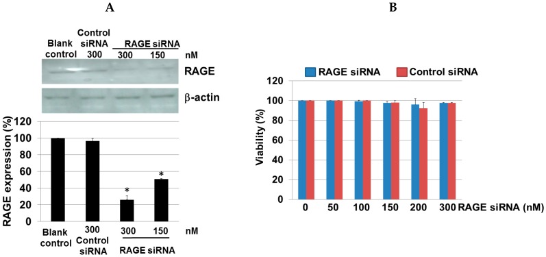
Figure 7.
Efficiency of RAGE gene knockdown in gingival epithelial cells. (A) A representative Western blot using antibodies against RAGE and β-actin is shown. The results showed decreased expression of RAGE protein in the siRNA group compared to the control siRNA and blank control groups. The intensity of the protein bands in Western blotting was quantified by densitometry and normalized to β-actin. Three independent measurements were performed. Data are expressed as mean ± SD. * p < 0.001 vs. the control group; (B) Cytotoxicity of the siRNA complexes was assessed by MTT assay. There was no evidence of cell toxicity found in the RAGE siRNA transfected cells. Data are expressed as the mean ± SD. Error bars indicate SD of at least three independent determinations.

