Figure 7.
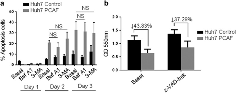
Effect of 3-MA, bafilomycin A1 and z-VAD-fmk on PCAF-induced apoptosis. (a) Huh7 PCAF cells and Huh7 Control cells in presence or absence of 5 mM 3-MA or 50 nM bafilomycin A1 (Baf A1) for 1–3 days, and then the Annexin V-FITC/PI double-staining analysis was performed. Cells in early apoptotic and necrotic/late apoptotic phases were quantified as apoptotic cells. The experiments were repeated at least three times. (b) Huh7 PCAF cells and Huh7 Control cells in presence or absence of 100 nM z-VAD-fmk for 2 days. Cytotoxicity was assessed by MTT assay. The experiments were repeated at least three times

