Figure 5.
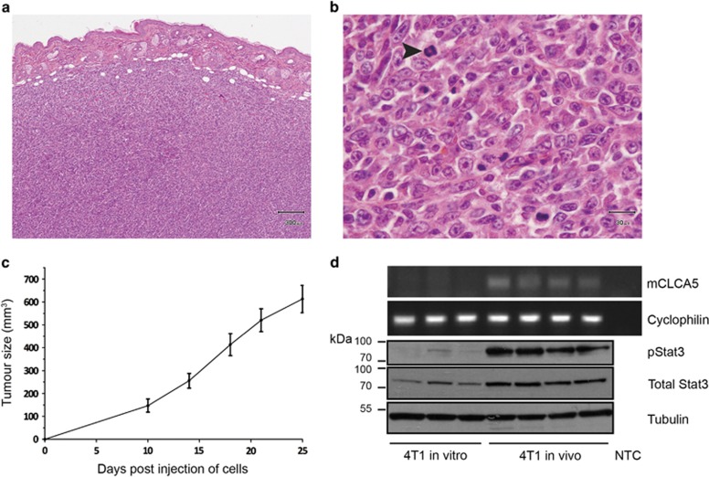
mCLCA5 is expressed in tumours resulting from implantation of 4T1 cells. (a and b) Representative photomicrographs demonstrating that orthotopic tumours derived from 4T1 cells are densely cellular, with minimal stroma and a moderately to markedly pleomorphic phenotype with numerous mitoses. Haematoxylin and eosin stain. Scale bar=300 μm (a) and 30 μm (b). Arrowhead indicates a mitotic figure. Several mitoses are present in this field. (c) Average tumour size in mm3 for tumours derived from injection of 1 × 10⁵ 4T1 cells in the mammary fat pad. Values are mean±S.D. from four biological repeats. (d) RT-PCR analysis for mCLCA5 and cyclophilin A, and western blot analysis of phosphorylated Stat3 (pStat3), total Stat3 and tubulin expression in 4T1 cells maintained in culture (4T1 in vitro) and in tumours resulting from implantation of 4T1 cells into the mammary fat pad of syngeneic mice (4T1 in vivo). Three independent biological repeats are shown for 4T1 in vitro; for 4T1 implanted in mice, four tumours from separate individuals are represented. NTC, no template control

