Figure 2.
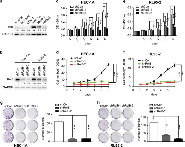
RelB depletion inhibits HEC-1A and RL95-2 cell growth. (a) RelB protein levels in different human EEC cells. (b) The protein level of RelB in HEC-1A and RL95-2 cells expressing shCon, shRelB-1 or shRelB-2. (c and e) Cell counting assay using the CCK-8 kit of HEC-1A (c) and RL95-2 (e) cells expressing shCon and shRelB-1 and -2. RelB knockdown significantly reduced cell proliferation. In all, 1 × 10³ cells per well were seeded in a 96-well plate, and cell viability was measured as the optical density at 450 nm (OD450) each day for 6 days. (d and f) The growth curve of HEC-1A (d) and RL95-2 (f) cells with shCon, shRelB-1, or shRelB-2. In total, 1 × 10⁴ cells per well were seeded in a 12-well plate, and cell numbers were assessed each day for 6 days. (g and h) The colony formation assay of HEC-1A (g) and RL95-2 (h) cells expressing shCon, shRelB-1 or shRelB-2. Colonies were counted after 10 days. RelB inhibition reduced the colony formation ability. All data are presented as the mean±S.D. of three independent experiments, *P<0.05, **P<0.01, ***P<0.001

