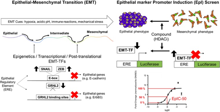
Figure 1.
Rationale of the EpI screen. EMT can be triggered by multifarious cues such as hypoxia, acidic pH, immune reactions, and mechanical stress. There are three states in the EMT: epithelial, intermediate, and mesenchymal. When a cancer cell undergoes EMT, alterations may occur at the epigenetic, transcriptional, and post-translational levels. EpI screen searches for drugs that reverses the effects of EMT-TFs on epithelial regulator elements. When EMT-TFs, such as SNAIL and ZEB, are upregulated, they sit on E-boxes and decreases the expression of E-cadherin, an epithelial gene. In addition, when an EMT-TF, such as GRHL2, is downregulated and does not bind to its binding sites, epithelial genes such as ERBB3 is suppressed Thus, a suppression of epithelial genes is an indicator of EMT, transforming the epithelial phenotype into a mesenchymal phenotype of a cancer cell. If the suppression of epithelial genes is removed, mesenchymal cells are able to regain its epithelial traits. In the drug discovery pipeline of EpI screen, the incorporation of epithelial regulator elements with luciferase, a bioluminescent enzyme, enables the quantification of potency of drugs in reversing EMT. A logarithmic concentration graph is plotted to determine the concentrations of drugs (logM) utilized from a library of HDACi to induce fold change, normalized to DMSO, in E-cadherin EpI activity. The arrow of EpIC-50 indicates the concentration corresponding to a 50% of maximum fold change

