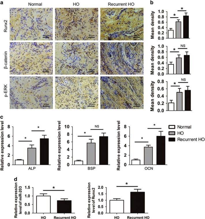
Figure 5.
Clinical HO specimens exhibit the overexpression of Runx2 and activated β-catenin and ERK signaling. (a) Normal bones (n=15) and primary (n=30) and recurrent HO tissues (n=7) were decalcified. The expression of Runx2, β-catenin and p-ERK was assayed by immunohistochemistry. (b) Quantification of Runx2, β-catenin and p-ERK expression. (c) Total RNA was isolated from normal bones and primary and recurrent HO tissues. The expression of ALP, BSP and OCN was examined with real-time PCR assays. (d) Total RNA was isolated from primary HO and recurrent HO tissues. The relative miR-203 and Runx2 levels were detected by real-time PCR assays. *P<0.01; NS, not significant

