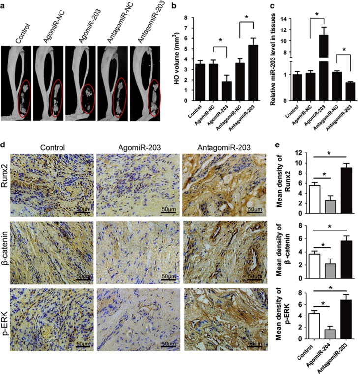
Figure 6.
Therapeutic overexpression of miR-203 inhibits HO in vivo. (a) The Achilles tendon of the mice was divided at its midpoint with a surgical knife to generate a traumatic HO model. Then the mice were injected weekly at the lesion with agomiR-203, antagomiR-203, or their NCs (PBS was used as the control). Representative micro-CT reconstructed images of HO in the control, agomiR-NC, agomiR-203, antagomiR-NC and antagomiR-203 mice. (b) Quantification of the HO volumes. (c) Total RNA was isolated from the mice HO. The relative miR-203 levels were detected by real-time PCR assays. (d) HO tissues from the mice were decalcified, and the expression of Runx2, β-catenin and p-ERK was detected by immunohistochemistry at week 8. (e) Quantification of Runx2, β-catenin and p-ERK expression. n=10 per group. The data are shown as the means±S.D. *P<0.01

