Figure 4.
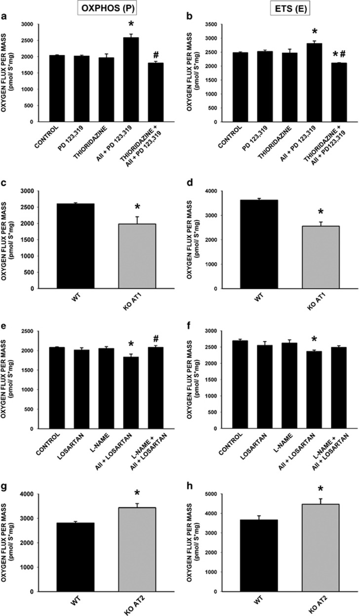
Effect of mitochondrial angiotensin receptors on mitochondrial respiration. (a and b) Activation of mitochondrial AT1 receptors with AII (i.e. AII+ PD123,319) induces an increase in both oxidative phosphorylation (P) and maximum respiratory rate (E), which was inhibited by pre-treatment of isolated mitochondria with the NOX4 inhibitor thioridazine (n=3–8). (c and d) Knockout mice for AT1 receptors (KO AT1; n=5) show lower mitochondrial respiration rates compared with wild-type littermate controls (WT). (e and f) Activation of mitochondrial AT2 receptors by AII (i.e. AII+losartan) produces a significant decrease in activated respiration (OXPHOS, P) and maximum respiration rate (maximum electron transport system, ETS, E) associated with complex I, which was blocked by pre-incubation of isolated mitochondria with the nitric oxide synthase (NOS) inhibitor, Nω-nitro-l-arginine methyl ester hydrochloride (l-NAME; n=5–8). (g and h) Mice lacking AT2 receptors (KO AT2; n=5) show an increased respiratory activity compared with wild-type mice (WT). Data are mean±S.E.M. *P<0.05 compared with control. #P<0.05 compared with the group treated with AII. One-way analysis of variance (ANOVA) and Bonferroni post hoc test (a, b, e, f) and Student's t-test(c, d, g, h)

