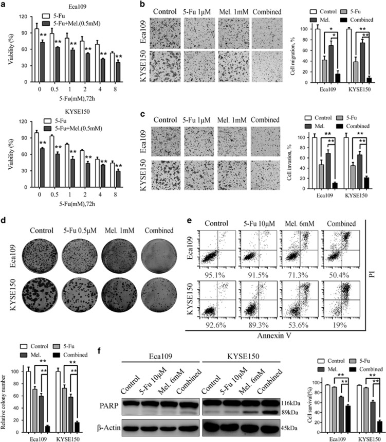
Figure 5.
Synergistic effects between melatonin and 5-Fu in ESCC cells in vitro. (a) Cell viability of Eca109 (upper panel) and KYSE150 (lower panel) cells treated with 5-Fu alone or combined with melatonin (0.5 mM) at indicated concentrations was detected by MTS. (b) Representative images (left panel) and quantification (right panel) of migration assays in Eca109 and KYSE150 cells treated with 5-Fu (1 μM) and melatonin (1 mM) for 24 h. (c) Representative images (left panel) and quantification (right panel) of invasion assays in the indicated cells treated with 5-Fu (1 μM) and melatonin (1 mM) for 24 h. (d) Representative images (upper panel) and quantification (lower panel) of colony formation in Eca109 and KYSE150 cells treated with 5-Fu (0.5 μM) and melatonin (1 mM) for 14 days. (e) Representative images (upper panel) and quantification (lower panel) of Annexin-V/PI assays in the indicated cells treated with 5-Fu (10 μM) and melatonin (6 mM) for 24 h. (f) Immunoblotting of PARP in Eca109 and KYSE150 cells treated with 5-Fu (10 μM) and melatonin (6 mM) for 24 h. β-Actin was used as a loading control. Data in (a), (b), (c), (d) and (e) are presented as mean±S.E. derived from three individual experiments with triplicate wells. *P<0.05 and **P<0.01 versus corresponding control. Error bars, S.E.

