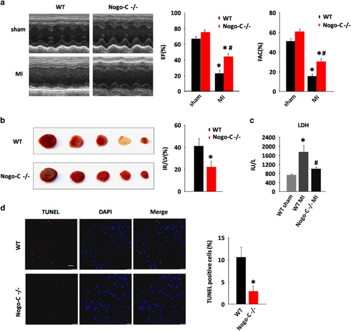
Figure 4.
Nogo-C knockout protects mouse heart from MI injury. (a) Typical example of M-mode echocardiograms of wildtype and Nogo-C−/− mouse hearts with (MI) or without (sham) LAD ligation for 24 h (left), and average data of EF (middle) and FAC (right). n=6 mice for each group. *P<0.05 versus sham wildtype mice; #P<0.05 versus MI wildtype mice. (b) Representative triphenyltetrazolium chloride (TTC) staining images of sequential heart sections (left), and the average data of TTC infarction size (right) of WT and Nogo-C−/− mice after LAD ligation for 24 h. n=4 mice,*P<0.05 versus wildtype mice. (c) Serum LDH activities of wildtype and Nogo-C−/− mice with or without LAD ligation for 24 h. n=5–11 mice. *P<0.05 versus WT sham; #P<0.05 versus WT MI. (d) TUNEL staining showing apoptotic cells in the border zone of MI from WT and Nogo-C−/− mouse hearts 24 h after LAD ligation (left), and the average data (right). n=4 mice. Scale bar=25 μm. *P<0.05 versus wildtype mice

