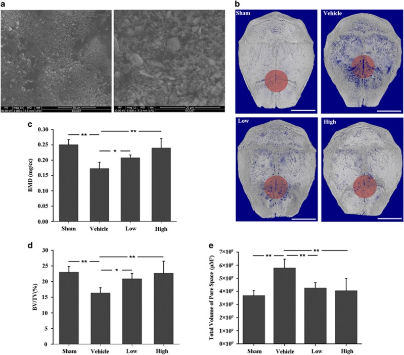
Figure 1.
DFO alleviated UHMWPE particles-induced mouse calvaria osteolysis. (a) Scanning electron micrograph of UHMWPE particles. (b) Representative micro-CT three-dimensional reconstructed images from each group. Scale bars, 3 mm. (c–e) BMD, BV/TV and total volume of pore space in the region of interest were measured. Low and high represent 10 and 30 mg/kg DFO application, respectively. n=6, *P<0.05, **P<0.01

