
Fig. 2.
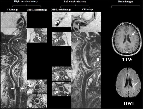
One patient with multiple co-existing intra- and extra-cranial plaques and recurrent stroke. The old infarct showed hypointense on brain T1W image (arrow) and the acute infarct showed hyperintense on brain DWI image (arrow). The multiple plaques can be seen on both left and right curved reconstructed (CR) images of MERGE. Seven plaques (hollow arrows) were identified on axial images after multiple planner reconstruction (MPR) in different cerebral artery segments