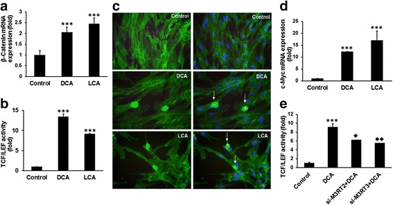
Fig. 5.

Bile acids induce Wnt-β-catenin signaling pathways and increase the expression of the target gene c-Myc in HCoEpiC. Real-time qPCR showing an increased expression of β-catenin mRNA in HCoEpiC cells following 72-h incubation in the presence of 100 μM DCA or LCA (a). Induction of transcriptional activity of TCF/LEF in HCoEpiC in response to 100 μM DCA or LCA treatments for 72 h (b). Photomicrographs showing increased nuclear localization of β-catenin in HCoEpiC following 72 h incubation with 100 μM DCA or LCA; controls were incubated with an equivalent volume of the vehicle (c): left panel , β-catenin immunostained cells; right panel, merged photograph of β-catenin and nucleus stained with DAPI; arrow, nuclear localization of β-catenin in cells. Increased expression of c-Myc in HCoEpiC following 72-h exposure to 100 μM DCA or LCA (d). DCA/LCA-mediated induction of transcriptional activity of TCF/LEF is greatly suppressed in M3R-downregulated HCoEpiC (e). Cells were photographed on a 100 μm scale at a magnification of 400×. Results expressed as mean ± standard deviation of three separate experiments. *P < 0.05, **P < 0.01 and ***P < 0.001. DCA (deoxycholic acid), LCA (lithocholic acid), TCF/LEF (T-cell factor/lymphoid-enhancing factor). Diamonds represent significant reduction in M3R down-regulated cells in response to DCA compared to those without siM3R transfected cells
