Fig. 5.

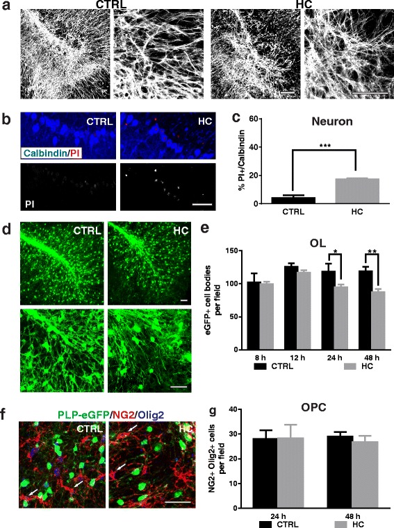
HC causes mild neuronal and mature oligodendroglial cell loss in mouse organotypic cerebellar slice culture. Immunostaining and quantitation of neuron and glia cell death in the slices treated with medium only (CTRL) or 10% HC for 48 h. a 25× (left panels) and 63× (right panels) objective images of slices stained with AST marker GFAP. b, c Calbindin-positive Purkinje neurons show increased PI staining in the presence of HC. d, e PLP-eGFP-positive OLs are mildly reduced in number following HC treatment for 48 h. 25× (upper) and 63× (lower) objective images of PLP-eGFP in slices treated for 48 h (d). eGFP+ cell bodies were quantified at 8, 12, 24, and 48 h in multiple 20× fields (e). f, g Slices were treated for 48 h and co-stained with Olig2 and NG2. Arrows mark Olig2+NG2+ OPCs. OPCs were counted at 24 or 48 h following HC administration. Statistical analyses were performed by multiple unpaired Student’s t test. *p < 0.05, **p < 0.01, n ≥ 3. Scale bars 50 μm
