Fig. 7.

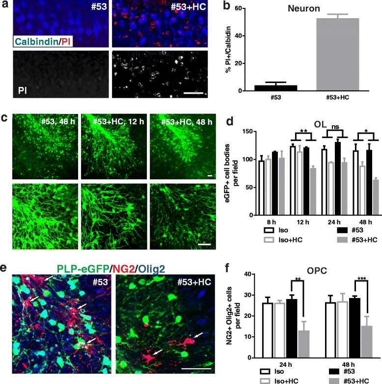
Increased neuron and OL death following astrocyte damage in brain slices. Immunostaining and quantification of neuron and oligodendrocyte cell death in the slices treated with rAb NMO#53 or isotype control rAb with or without 10% HC for 48 h. PI-stained cells double stained with Purkinje neuron marker Calbindin (a). Quantification of PI-labeled cells co-localized with Calbindin, as a percent of the total Calbindin+ cells (b). 25× (upper panel) and 63× (lower panel) objective images of PLP-eGFP in slices (c). Quantification of eGFP+ OLs in slices at 8, 12, 24, and 48 h after treatment (d). Cerebellar slices treated for 24 h with rAb #53 and HC were co-stained with Olig2 and NG2 to identify OPCs (e). Arrows mark OPCs. f OPCs were counted in slices treated for 24 and 48 h. Iso: negative isotope control rAb. Statistical analyses were performed by multiple unpaired Student’s t test for single comparison (f) or by two-way ANOVA for grouped comparisons (d). *p < 0.05, **p < 0.01, ***p < 0.001, ns not significant, n ≥ 3. Scale bars 50 μm
