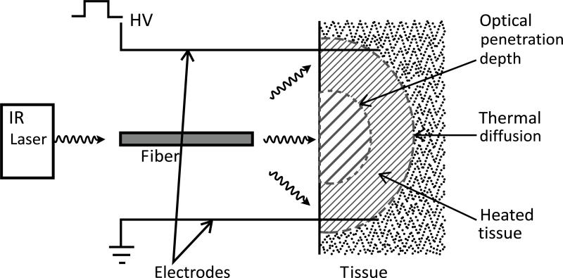
Fig. (1).
TAGET Device. Schematic of an optical fiber that was connected to an infrared laser and inserted into the device. The fiber is situated centrally between 4 electrodes. The fiber is also placed 1 cm above a 3 mm opening. This gap and opening allows for an increased spot size of the light as it exits into the space between the opening and the tissue which is set at 5 mm.

