Fig. 4.

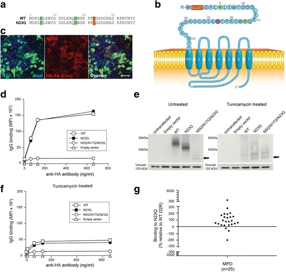
Point mutation of D2R third N-glycosylation site does not abolish anti-D2R antibody binding. a Alignment of extracellular N-terminal 37 amino acids of N23Q and WT D2R. Green = putative N-glycosylation sites; Red = N to Q point mutation. b Schematic of N23Q at cell surface. Red = 3xHA tag; Green = N-glycosylation site; Blue = D2R sequence; Purple = point mutation. c, d Confocal images and flow cytometry analysis after live immunolabeling of WT D2R and N23Q-transfected HEK293 cells with an anti-HA antibody showed a similar level of surface expression for both constructs (scale bar = 50 μm). No D2R expression from empty vector was observed. Reduced surface expression of N5Q/N17Q/N23Q was observed. Representative data out of three independent experiments is shown. e Loss of N-linked glycan and decrease in molecular weight was detected by western blot analysis in untreated (left panel) and tunicamycin-treated (right panel) WT D2R-, N23Q-, and N5Q/N17Q/N23Q-transfected HEK293 cells. Tunicamycin treatment led to blockade of glycosylation in all mutant-transfected cells. Representative data out of three experiments is shown. f Deglycosylation led to impairment in trafficking and decreased cell surface expression of WT D2R, N23Q, and N5Q/N17Q/N23Q. g Sera from anti-D2R antibody-positive movement and psychiatric disorders (MPD, n = 25) were incubated with empty vector-, WT D2R-, and N23Q-transfected live HEK293 cells at 1:50 dilution, followed by AF647-conjugated anti-human IgG secondary antibody, and flow cytometry analysis. Percentage of sera binding to N23Q (MFI %) was calculated using the formula described in Material and Methods. 88% patients (22/25) recognized N23Q, whereas 12% (3/25) showed no immunoreactivity to N23Q. Binding threshold is represented by solid line on graph. Representative data out of three experiments is shown
