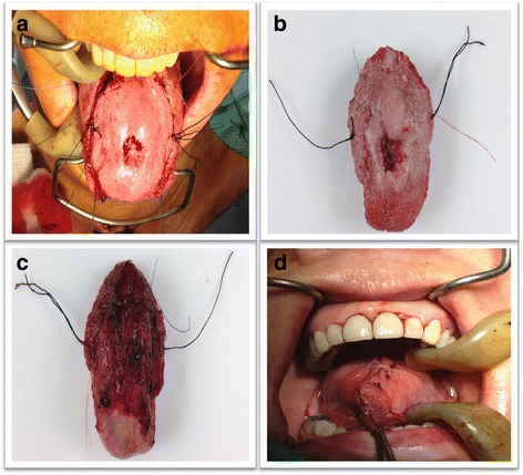
Fig. 3.

Surgical specimen from resection. Intraoperative Mohs margins. Image (a) demonstrates the surgical specimen about to be resected with proper orientation in both patient and the specimen. Images (b and c) demonstrate the resected specimen with corresponding sutures. Image (d) demonstrates closing time at the end of the operation once all margins were negative
