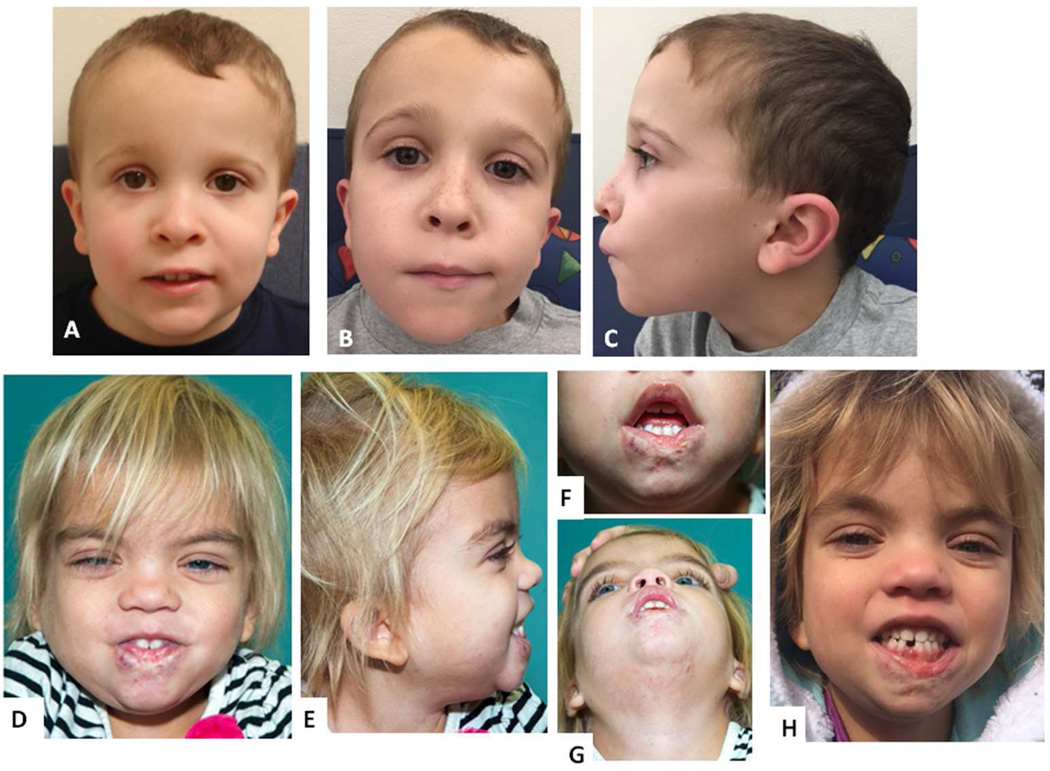
Figure 2.
Facial photographs of Patient 2 at ages 5 5/12 years (A) and 9 years (B, C), note low-set, posteriorly angulated ears; facial photographs of Patient 3 at age 4 years, note hypertelorism and epicanthus, fine blond hair, low-set ears (D, E), scarring of her lower lip after hemangioma treatment (F, G), and at age 5 years (H).

